Artlabeling Activity Organization of the Sympathetic Division of the Ans
Learning Objectives
- Name the components that generate the sympathetic and parasympathetic responses of the autonomic nervous system
- Explain the differences in output connections inside the two divisions of the autonomic nervous system
- Draw the signaling molecules and receptor proteins involved in advice within the ii divisions of the autonomic nervous system
The nervous system tin exist divided into 2 functional parts: the somatic nervous arrangement and the autonomic nervous system. The major differences between the ii systems are evident in the responses that each produces. The somatic nervous system causes contraction of skeletal muscles. The autonomic nervous system controls cardiac and smooth muscle, equally well as glandular tissue. The somatic nervous system is associated with voluntary responses (though many can happen without conscious sensation, like animate), and the autonomic nervous arrangement is associated with involuntary responses, such every bit those related to homeostasis.
The autonomic nervous system regulates many of the internal organs through a residual of two aspects, or divisions. In ition to the endocrine system, the autonomic nervous system is instrumental in homeostatic mechanisms in the body. The two divisions of the autonomic nervous organisation are thesympathetic division and theparasympathetic division. The sympathetic system is associated with thefight-or-flight response, and parasympathetic activity is referred to by the epithet ofrest and digest. Homeostasis is the residue between the two systems. At each target effector, dual innervation determines action. For instance, the center receives connections from both the sympathetic and parasympathetic divisions. Ane causes heart charge per unit to increment, whereas the other causes centre charge per unit to decrease.
Watch this video to larn more nigh adrenaline and the fight-or-flying response.
When someone is said to take a rush of adrenaline, the image of bungee jumpers or skydivers usually comes to listen. Just adrenaline, also known equally epinephrine, is an of import chemical in analogous the body's fight-or-flight response. In this video, y'all look within the physiology of the fight-or-flight response, as envisioned for a fireman. His trunk's reaction is the event of the sympathetic division of the autonomic nervous system causing system-wide changes equally it prepares for extreme responses. What 2 changes does adrenaline bring about to aid the skeletal musculus response?
Sympathetic Sectionalisation of the Autonomic Nervous System
To answer to a threat—to fight or to run away—the sympathetic system causes divergent furnishings as many different effector organs are activated together for a mutual purpose. More oxygen needs to be inhaled and delivered to skeletal muscle. The respiratory, cardiovascular, and musculoskeletal systems are all activated together. itionally, sweating keeps the backlog heat that comes from muscle wrinkle from causing the body to overheat. The digestive organization shuts down and then that blood is not absorbing nutrients when it should be delivering oxygen to skeletal muscles. To coordinate all these responses, the connections in the sympathetic system diverge from a limited region of the central nervous system (CNS) to a wide assortment of ganglia that project to the many effector organs simultaneously. The complex ready of structures that compose the output of the sympathetic system make it possible for these disparate effectors to come together in a coordinated, systemic modify.
The sympathetic partitioning of the autonomic nervous organisation influences the diverse organ systems of the body through connections emerging from the thoracic and upper lumbar spinal string. It is referred to every bit thethoracolumbar arrangement to reflect this anatomical basis. Acentral neuron in the lateral horn of whatsoever of these spinal regions projects to ganglia adjacent to the vertebral column through the ventral spinal roots.
The majority of ganglia of the sympathetic organization belong to a network ofsympathetic chain ganglia that runs alongside the vertebral column. The ganglia appear as a series of clusters of neurons linked by axonal bridges. There are typically 23 ganglia in the chain on either side of the spinal column. Three correspond to the cervical region, 12 are in the thoracic region, four are in the lumbar region, and iv correspond to the sacral region. The cervical and sacral levels are non connected to the spinal cord straight through the spinal roots, merely through ascending or descending connections through the bridges within the chain.
A diagram that shows the connections of the sympathetic system is somewhat like a circuit diagram that shows the electrical connections between dissimilar receptacles and devices. In Effigy 1, the "circuits" of the sympathetic organisation are intentionally simplified.

Effigy 1. Connections of Sympathetic Segmentation of the Autonomic Nervous System. Neurons from the lateral horn of the spinal cord (preganglionic neurons) project to the concatenation ganglia on either side of the vertebral column or to collateral (prevertebral) ganglia that are anterior to the vertebral column in the intestinal cavity. Axons from these ganglionic neurons (postganglionic fibers) then projection to target effectors throughout the body.
To proceed with the illustration of the excursion diagram, in that location are three different types of "junctions" that operate within the sympathetic system (Figure two). The first blazon is most directly: the sympathetic nerve projects to the chain ganglion at the aforementioned level as thetarget effector (the organ, tissue, or gland to be innervated).
An instance of this type is spinal nerve T1 that synapses with the T1 chain ganglion to innervate the trachea. The fibers of this co-operative are calledwhite rami communicantes (atypical = ramus communicans); they are myelinated and therefore referred to equally white (see Figure 2a). The axon from the central neuron (the preganglionic fiber shown as a solid line) synapses with theganglionic neuron (with the postganglionic cobweb shown equally a dashed line). This neuron then projects to a target effector—in this case, the trachea—viagray rami communicantes, which are unmyelinated axons.
In some cases, the target effectors are located superior or inferior to the spinal segment at which the preganglionic fiber emerges. With respect to the "wiring" involved, the synapse with the ganglionic neuron occurs at chain ganglia superior or junior to the location of the central neuron. An example of this is spinal nervus T1 that innervates the eye. The spinal nerve tracks upwards through the concatenation until it reaches thesuperior cervical ganglion, where it synapses with the postganglionic neuron (run across Effigy 2b). The cervical ganglia are referred to asparavertebral ganglia, given their location adjacent to prevertebral ganglia in the sympathetic concatenation.
Not all axons from the central neurons terminate in the chain ganglia. itional branches from the ventral nerve root continue through the chain and on to one of the collateral ganglia equally thegreater splanchnic nervus or lesser splanchnic nerve. For example, the greater splanchnic nervus at the level of T5 synapses with a collateral ganglion outside the chain before making the connection to the postganglionic nerves that innervate the stomach (see Figure 2c).
Collateral ganglia, besides calledprevertebral ganglia, are situated anterior to the vertebral column and receive inputs from splanchnic nerves also as central sympathetic neurons. They are associated with decision-making organs in the abdominal cavity, and are too considered function of the enteric nervous system. The iii collateral ganglia are theceliac ganglion, thesuperior mesenteric ganglion, and thejunior mesenteric ganglion (see Figure 1). The word celiac is derived from the Latin word "coelom," which refers to a body cavity (in this example, the abdominal cavity), and the word mesenteric refers to the digestive system.

Effigy 2. Sympathetic Connections and Chain Ganglia. The axon from a central sympathetic neuron in the spinal cord can project to the periphery in a number of unlike ways. (a) The fiber can project out to the ganglion at the aforementioned level and synapse on a ganglionic neuron. (b) A branch can project to more than superior or junior ganglion in the chain. (c) A branch can project through the white ramus communicans, but non terminate on a ganglionic neuron in the chain. Instead, it projects through one of the splanchnic fretfulness to a collateral ganglion or the adrenal medulla (not pictured).
An axon from the central neuron that projects to a sympathetic ganglion is referred to as apreganglionic fiber or neuron, and represents the output from the CNS to the ganglion. Because the sympathetic ganglia are side by side to the vertebral cavalcade, preganglionic sympathetic fibers are relatively short, and they are myelinated. Apostganglionic cobweb—the axon from a ganglionic neuron that projects to the target effector—represents the output of a ganglion that directly influences the organ.
Compared with the preganglionic fibers, postganglionic sympathetic fibers are long because of the relatively greater distance from the ganglion to the target effector. These fibers are unmyelinated. (Annotation that the term "postganglionic neuron" may exist used to describe the project from a ganglion to the target. The problem with that usage is that the cell body is in the ganglion, and only the fiber is postganglionic. Typically, the term neuron applies to the entire cell.)
One type of preganglionic sympathetic fiber does not terminate in a ganglion. These are the axons from central sympathetic neurons that project to theadrenal medulla, the interior portion of the adrenal gland. These axons are withal referred to as preganglionic fibers, but the target is non a ganglion. The adrenal medulla releases signaling molecules into the bloodstream, rather than using axons to communicate with target structures. The cells in the adrenal medulla that are contacted by the preganglionic fibers are chosenchromaffin cells. These cells are neurosecretory cells that develop from the neural crest along with the sympathetic ganglia, reinforcing the idea that the gland is, functionally, a sympathetic ganglion.
The projections of the sympathetic partition of the autonomic nervous system diverge widely, resulting in a broad influence of the system throughout the body. As a response to a threat, the sympathetic system would increase middle rate and breathing rate and crusade blood menses to the skeletal muscle to increment and blood menstruum to the digestive system to decrease. Sweat gland secretion should also increase as part of an integrated response.
All of those physiological changes are going to be required to occur together to run away from the hunting lioness, or the modern equivalent. This deviation is seen in the branching patterns of preganglionic sympathetic neurons—a unmarried preganglionic sympathetic neuron may have 10–20 targets. An axon that leaves a central neuron of the lateral horn in the thoracolumbar spinal cord will pass through the white ramus communicans and enter the sympathetic chain, where it volition branch toward a variety of targets. At the level of the spinal cord at which the preganglionic sympathetic fiber exits the spinal string, a branch will synapse on a neuron in the adjacent chain ganglion.
Some branches will extend up or down to a dissimilar level of the chain ganglia. Other branches volition pass through the chain ganglia and project through one of the splanchnic nerves to a collateral ganglion. Finally, some branches may project through the splanchnic nerves to the adrenal medulla. All of these branches mean that i preganglionic neuron can influence different regions of the sympathetic system very broadly, past interim on widely distributed organs.
Parasympathetic Division of the Autonomic Nervous Organisation
The parasympathetic sectionalization of the autonomic nervous system is named considering its cardinal neurons are located on either side of the thoracolumbar region of the spinal cord (para– = "beside" or "almost"). The parasympathetic system can also be referred to as thecraniosacral system (or outflow) because the preganglionic neurons are located in nuclei of the brain stalk and the lateral horn of the sacral spinal cord.
The connections, or "circuits," of the parasympathetic partition are similar to the general layout of the sympathetic division with a few specific differences (Effigy 3). The preganglionic fibers from the cranial region travel in cranial nerves, whereas preganglionic fibers from the sacral region travel in spinal nerves. The targets of these fibers are final ganglia, which are located near—or fifty-fifty within—the target effector. These ganglia are often referred to asintramural ganglia when they are institute within the walls of the target organ. The postganglionic fiber projects from the terminal ganglia a brusque altitude to the target effector, or to the specific target tissue within the organ. Comparison the relative lengths of axons in the parasympathetic organization, the preganglionic fibers are long and the postganglionic fibers are short because the ganglia are close to—and sometimes within—the target effectors.
The cranial component of the parasympathetic organisation is based in detail nuclei of the encephalon stem. In the midbrain, theEddinger–Westphal nucleus is part of the oculomotor complex, and axons from those neurons travel with the fibers in the oculomotor nerve (cranial nervus III) that innervate the extraocular muscles. The preganglionic parasympathetic fibers within cranial nervus Iii terminate in theciliary ganglion, which is located in the posterior orbit. The postganglionic parasympathetic fibers then project to the smooth muscle of the iris to control pupillary size. In the upper medulla, the salivatory nuclei incorporate neurons with axons that project through the facial and glossopharyngeal nerves to ganglia that control salivary glands. Tear product is influenced past parasympathetic fibers in the facial nerve, which activate a ganglion, and ultimately the lacrimal (tear) gland.
Neurons in thedorsal nucleus of the vagus nervus and thenucleus ambiguus projection through the vagus nerve (cranial nervus X) to the terminal ganglia of the thoracic and abdominal cavities. Parasympathetic preganglionic fibers primarily influence the heart, bronchi, and esophagus in the thoracic cavity and the stomach, liver, pancreas, gallbladder, and pocket-size intestine of the intestinal cavity. The postganglionic fibers from the ganglia activated by the vagus nervus are often incorporated into the construction of the organ, such as themesenteric plexus of the digestive tract organs and the intramural ganglia.

Figure 3.Connections of Parasympathetic Sectionalization of the Autonomic Nervous System Neurons from encephalon-stem nuclei, or from the lateral horn of the sacral spinal string, projection to terminal ganglia almost or inside the various organs of the trunk. Axons from these ganglionic neurons so project the short distance to those target effectors.
Chemical Signaling in the Autonomic Nervous Organisation
Where an autonomic neuron connects with a target, at that place is a synapse. The electrical signal of the action potential causes the release of a signaling molecule, which will bind to receptor proteins on the target prison cell. Synapses of the autonomic system are classified as eithercholinergic, meaning thatacetylcholine (ACh) is released, oradrenergic, meaning thatnorepinephrine is released. The terms cholinergic and adrenergic refer not only to the signaling molecule that is released but also to the course of receptors that each binds.
The cholinergic organization includes two classes of receptor: thenicotinic receptor and themuscarinic receptor. Both receptor types bind to ACh and cause changes in the target cell. The nicotinic receptor is aligand-gated cation channel and the muscarinic receptor is aG poly peptide–coupled receptor. The receptors are named for, and differentiated by, other molecules that bind to them. Whereas nicotine will bind to the nicotinic receptor, and muscarine volition bind to the muscarinic receptor, at that place is no cross-reactivity betwixt the receptors. The situation is similar to locks and keys.
Imagine two locks—i for a classroom and the other for an office—that are opened by two carve up keys. The classroom fundamental will not open up the office door and the role primal will not open the classroom door. This is like to the specificity of nicotine and muscarine for their receptors. Nonetheless, a master key tin open multiple locks, such as a primary key for the Biology Section that opens both the classroom and the function doors. This is similar to ACh that binds to both types of receptors. The molecules that define these receptors are not crucial—they are simply tools for researchers to utilize in the laboratory. These molecules areexogenous, meaning that they are made outside of the man body, so a researcher can use them without any misreckoningendogenous results (results caused past the molecules produced in the trunk).
The adrenergic organisation also has two types of receptors, named thealpha (α)-adrenergic receptor andbeta (β)-adrenergic receptor. Unlike cholinergic receptors, these receptor types are not classified past which drugs can bind to them. All of them are Grand poly peptide–coupled receptors. There are iii types of α-adrenergic receptors, termed αone, α2, and αthree, and there are two types of β-adrenergic receptors, termed βone and β2. An itional aspect of the adrenergic system is that at that place is a second signaling molecule calledepinephrine. The chemical difference betwixt norepinephrine and epinephrine is the ition of a methyl group (CH3) in epinephrine. The prefix "nor-" actually refers to this chemic difference, in which a methyl group is missing.
The term adrenergic should remind you of the word adrenaline, which is associated with the fight-or-flight response described at the beginning of the chapter. Adrenaline and epinephrine are two names for the same molecule. The adrenal gland (in Latin, advert– = "on top of"; renal = "kidney") secretes adrenaline. The ending "-ine" refers to the chemical beingness derived, or extracted, from the adrenal gland. A similar construction from Greek instead of Latin results in the word epinephrine (epi– = "to a higher place"; nephr– = "kidney"). In scientific usage, epinephrine is preferred in the United States, whereas adrenaline is preferred in Swell Britain, because "adrenalin" was once a registered, proprietary drug name in the United States. Though the drug is no longer sold, the convention of referring to this molecule by the two different names persists. Similarly, norepinephrine and noradrenaline are two names for the same molecule.
Having understood the cholinergic and adrenergic systems, their role in the autonomic system is relatively simple to understand. All preganglionic fibers, both sympathetic and parasympathetic, release ACh. All ganglionic neurons—the targets of these preganglionic fibers—have nicotinic receptors in their cell membranes. The nicotinic receptor is a ligand-gated cation aqueduct that results in depolarization of the postsynaptic membrane. The postganglionic parasympathetic fibers likewise release ACh, just the receptors on their targets are muscarinic receptors, which are Grand protein–coupled receptors and do non exclusively crusade depolarization of the postsynaptic membrane. Postganglionic sympathetic fibers release norepinephrine, except for fibers that projection to sweat glands and to blood vessels associated with skeletal muscles, which release ACh (Table ane).
| Table ane | ||
|---|---|---|
| Sympathetic | Parasympathetic | |
| Preganglionic | Acetylcholine > nicotinic receptor | Acetylcholine > nicotinic receptor |
| Postganglionic | Norepinephrine > a or B-adrenergic receptors Acetylcholine > muscarinic receptor (associated with sweat glands and the blood vessels associated with skeletal muscles only | Acetylcholine > muscarinic receptor |
Signaling molecules can belong to ii broad groups. Neurotransmitters are released at synapses, whereas hormones are released into the bloodstream. These are simplistic definitions, merely they tin help to clarify this point. Acetylcholine can exist considered a neurotransmitter because it is released past axons at synapses. The adrenergic system, however, presents a challenge. Postganglionic sympathetic fibers release norepinephrine, which can be considered a neurotransmitter. But the adrenal medulla releases epinephrine and norepinephrine into circulation, then they should be considered hormones.
What are referred to here every bit synapses may not fit the strictest definition of synapse. Some sources will refer to the connectedness between a postganglionic cobweb and a target effector as neuroeffector junctions; neurotransmitters, as divers above, would be called neuromodulators. The structure of postganglionic connections are not the typical synaptic finish bulb that is found at the neuromuscular junction, simply rather are chains of swellings along the length of a postganglionic fiber called avaricosity (Figure 4).
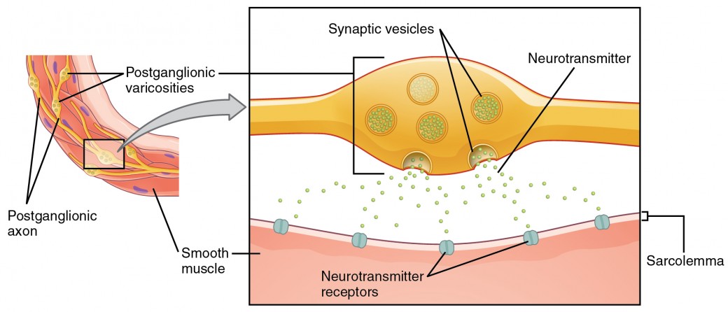
Figure 4. Autonomic Varicosities. The connection betwixt autonomic fibers and target effectors is non the same as the typical synapse, such as the neuromuscular junction. Instead of a synaptic end bulb, a neurotransmitter is released from swellings along the length of a fiber that makes an extended network of connections in the target effector.
Everyday Connections:Fight or Flying? What Virtually Fright and Freeze?
The original usage of the epithet "fight or flight" comes from a scientist named Walter Cannon who worked at Harvard in 1915. The concept of homeostasis and the performance of the sympathetic system had been introduced in France in the previous century. Cannon expanded the idea, and introduced the idea that an animal responds to a threat past preparing to stand up and fight or run away. The nature of this response was thoroughly explained in a book on the physiology of pain, hunger, fright, and rage.
When students learn about the sympathetic system and the fight-or-flight response, they often end and wonder about other responses. If yous were faced with a lioness running toward you lot equally pictured at the beginning of this chapter, would yous run or would you stand your ground? Some people would say that they would freeze and not know what to practise. So isn't in that location really more to what the autonomic organisation does than fight, flying, rest, or assimilate. What about fearfulness and paralysis in the face of a threat?
The common epithet of "fight or flight" is being enlarged to be "fight, flight, or fear" or even "fight, flight, fearfulness, or freeze." Cannon's original contribution was a catchy phrase to express some of what the nervous organisation does in response to a threat, merely it is incomplete. The sympathetic arrangement is responsible for the physiological responses to emotional states. The proper noun "sympathetic" can be said to mean that (sym– = "together"; –pathos = "pain," "suffering," or "emotion").
Sentinel this video to learn more than about the nervous organisation.
Equally described in this video, the nervous organization has a way to bargain with threats and stress that is split from the witting control of the somatic nervous system. The system comes from a time when threats were near survival, but in the mod age, these responses get office of stress and anxiety. This video describes how the autonomic system is just role of the response to threats, or stressors. What other organ system gets involved, and what part of the brain coordinates the ii systems for the entire response, including epinephrine (adrenaline) and cortisol?
Source: https://courses.lumenlearning.com/ap1/chapter/divisions-of-the-autonomic-nervous-system/
0 Response to "Artlabeling Activity Organization of the Sympathetic Division of the Ans"
Post a Comment